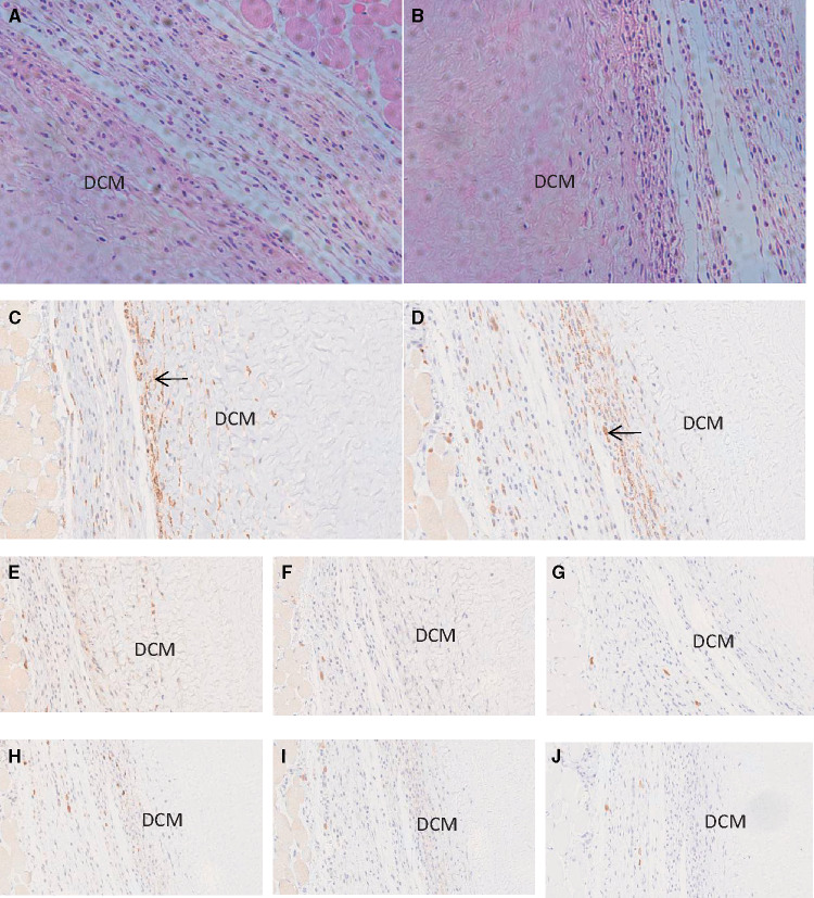
Figure 4.
HE and immunohistochemical staining of infiltrating cells in decellularized corneal matrix after 4 weeks of implantation in the WT mice and GTKO mice. Figures of (A) HE staining, (C) CD68+, (E) CD3+, (F) CD4+, and (G) CD8+ were obtained from WT mice; (B) HE staining, (D) CD68+, (H) CD3+, (I) CD4+, and (J) CD8+ were images from GTKO mice. The black arrows showed the positive staining cells. DCM: decellularized corneal matrix.

