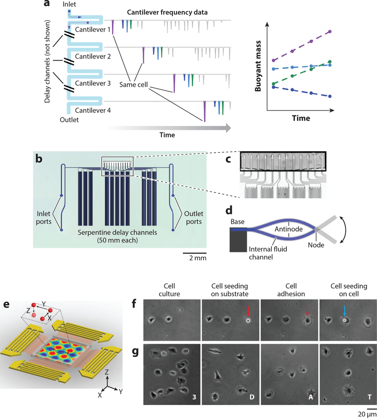
Figure 3.
Single-cell analysis via acoustic microfluidics. (a–d) Design and implementation of the serial suspended microchannel resonator (SMR) array. (a) Schematic working mechanism of serial SMR array. The mass accumulation rate of single cells flowing through can be monitored with respect to time. (b) Device layout and (c) microscopic picture of the serial SMR array. The serpentine channels create a time delay for cells to grow. (d) Schematic side view of a suspended microchannel vibrating in the second bending mode. (e–g)3D acoustic tweezers. (e) Schematic device layout. A particle can be dynamically programmed to move in 3D space in the microchamber. (f) Single-cell printing. (g) Creating arbitrary cell adhesion patterns via 3D acoustic tweezers. Panels a–d adapted with permission from Reference 115. Copyright 2016, Springer Nature. Panels e–g adapted with permission from Reference 144 and the National Academy of Sciences.

