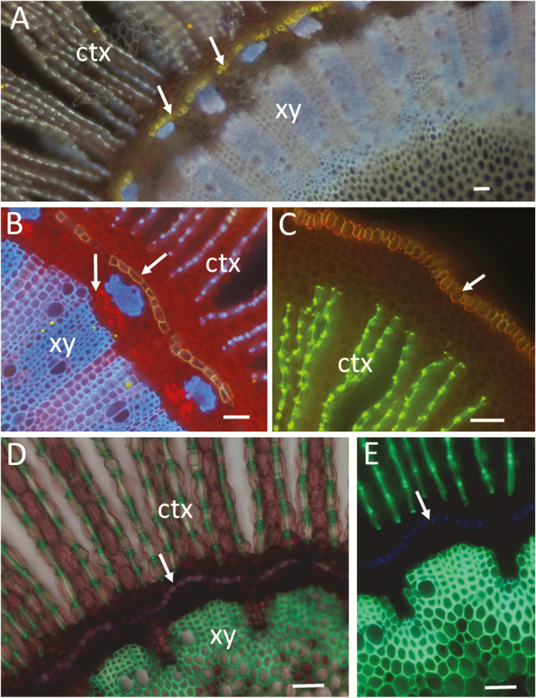
Figure 3.
Lignified, suberized and cellulosic walls in hand-cut transverse sections of root of a black mangrove plant (Bruguiera gymnorrhiza visualized by wide-field fluorescence (Olympus BX60). (A) Fluorol yellow staining for visualization of suberized endodermis (arrows). Inner cortex (ctx), and xylem (xy) are visualized with the blue autofluorescence of lignified walls (340–380 nm excitation, LP 420 emission). (B) Fluorol yellow/Congo red staining showing suberized endodermis (inclined arrow) and cellulosic walls of cortical and phloem cells (red). Note the stronger red fluorescence from phloem (vertical arrow). Xylem, phloem sclerenchyma and lignified thickenings of cortical parenchyma cells are visualized with the blue autofluorescence of lignin (340–380 nm excitation, LP 420 emission). (C) Fluorol yellow/acridine orange staining showing suberized epidermis (arrow) and lignified thickenings of cortical parenchyma cells (green). Merged green and red channels (BP 510–530; BP 590–610). (D) Acridine orange staining showing lignified walls (green fluorescence) in xylem and cortex. A combination of transmitted white light and wide-field fluorescence (340–380 nm excitation, LP 420 emission) was used to show the parenchyma cells and the intercellular air spaces in the cortex. (E) The same section as in (D) but visualized only by fluorescence without the transmitted light. Note the blue autofluorescence of Casparian thickenings (arrow) in the endodermal cells that at an early stage of development are not stained with the acridine orange. Bars = 50 µm (A through E).

