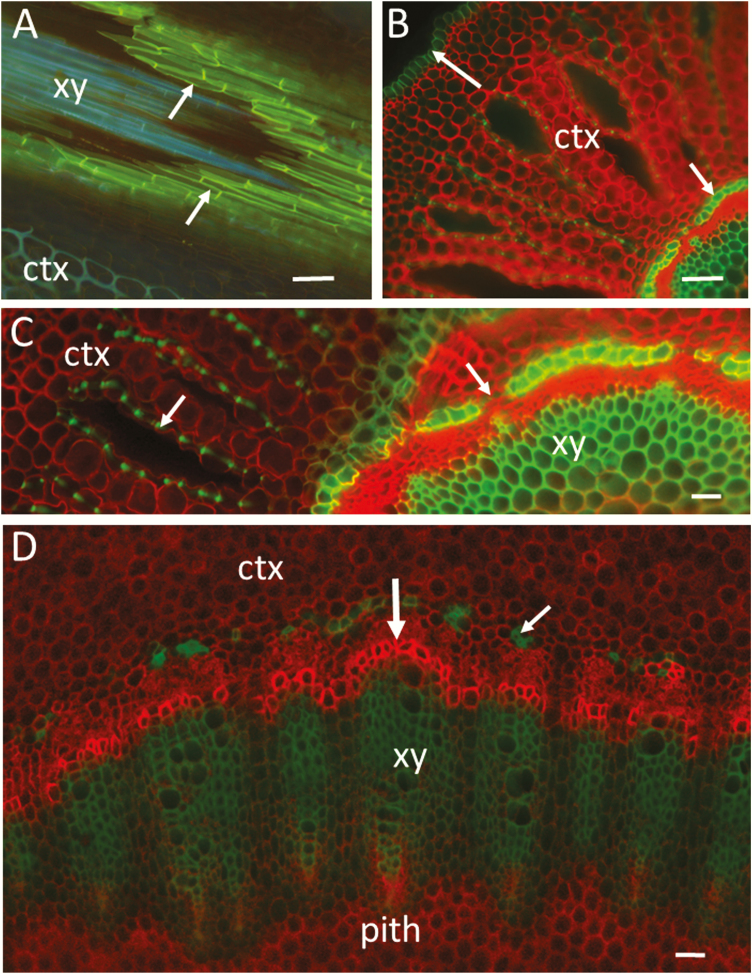
Figure 4.
Hand-cut sections of root and stem of a black mangrove plant (B. gymnorrhiza) visualized by wide-field fluorescence (Axio Scope A1; Carl Zeiss). (A) Longitudinal-oblique section through the root endodermis (arrows) showing xylem inside the endodermal cylinder. Staining with fluorol yellow (UV excitation and LP 420 emission). Suberized cell walls are yellow (or mixed blue-yellow) and lignified walls (xylem and cortex) emit blue-green autofluorescence. (B) Transverse section of a root showing the cortex, epidermis (upper arrow) and endodermis (lower arrow). Staining with Congo red and two-channel imaging (green ex/em: BP 450–490/BP 500–550, red ex/em: BP 539–563/BP 570–640). The composite image showing red staining of cellulosic walls and green autofluorescence of lignin and suberin. (C) Transverse section of a root showing inner cortex, endodermis, phloem and xylem. Note the green autofluorescence of lignified thickenings of cortical parenchyma cells (left arrow). The right arrow points to a transfusion endodermal cell with no suberin and lignin in the cell wall. The same staining and imaging conditions as in (B). (D) Transverse section through the hypocotyl of the same plant as in (C) showing inner cortex, xylem and small portion of the pith. Developing xylem cells with unlignified walls (vertical arrow) are stained stronger in bright red. The small arrow points to lignified cells on the outer side of the phloem. The same staining and imaging conditions as in (B). Bars = 100 µm (A and B), 50 µm (C and D). ctx, cortex; xy, xylem.

