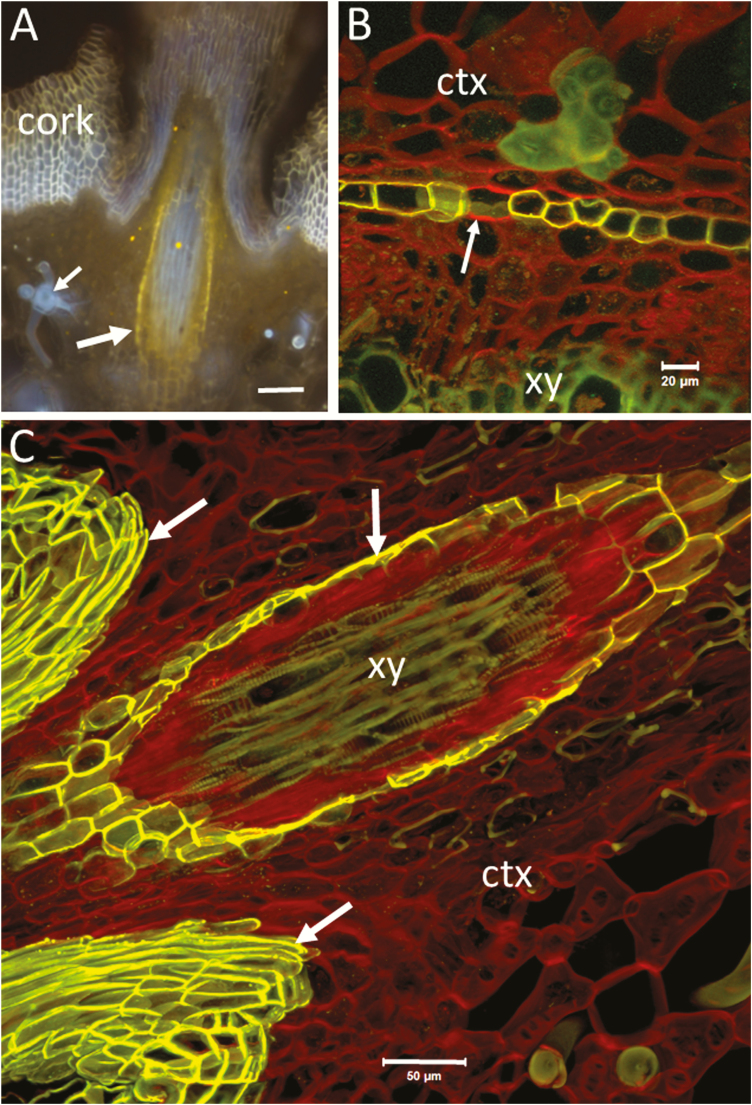
Figure 5.
Hand-cut transverse sections of roots of a Rhizophora apiculata plant visualized after staining with fluorol yellow (A) and after double staining with fluorol yellow/Congo red (B and C). The image in (A) was obtained by wide-field fluorescence with UV excitation and LP 420 emission (Olympus BX60). The images in (B and C) are merged two channels by CLSM (Zeiss LSM510; ex/em: 458/BP 500–550; 488/LP 560). The green and red emission from fluorol yellow-stained suberized walls is considerably stronger than the green and red from lignified and non-lignified walls, respectively. The resulting image shows red staining of polysaccharides, yellow (overlapped green and red) for suberized cell walls and green for lignin. (A) A lateral rootlet (obliquely cut) shown at the outer region of the cortex of the mother root. The large arrow points to yellow-stained endodermis of the rootlet. Note the blue autofluorescence of xylem inside the endodermal cylinder of the rootlet. The blue and yellow fluorescence of exodermal tissue (cork) indicates presence of lignin and suberin. Blue fluorescence is also emitted from lignified astrosclereids in the cortex (small arrow). (B) A view of the endodermal region by CLSM (maximum projection of 47 optical sections at 0.7-µm intervals; C-Apochromat 40×/1.2 Water). Non-lignified cell walls are stained in red. The arrow points to a transfusion endodermal cell with no suberin or lignin in the cell wall. The autofluorescence of lignified walls of xylem and sclerenchymatic cells in the cortex is shown in green. (C) A similar rootlet as in (A) but visualized in more detail by CLSM (maximum projection of nine optical sections at 2.3-µm intervals; Plan-Apochromat 20×/0.75). The arrows on the left point to exodermis. The vertical arrow points to the endodermis of the rootlet. Note the autofluorescence of xylem inside the endodermal cylinder (green). Phloem and parenchymatic cells with unlignified walls (Congo red staining) occupy the space between the xylem and endodermis. Note also the loose arrangement of cellulosic parenchyma cells in the cortex of the mother root resulting in formation of intercellular air spaces. Bars = 100 µm (A), 20 µm (B) and 50 µm (C). ctx, cortex; xy, xylem.

