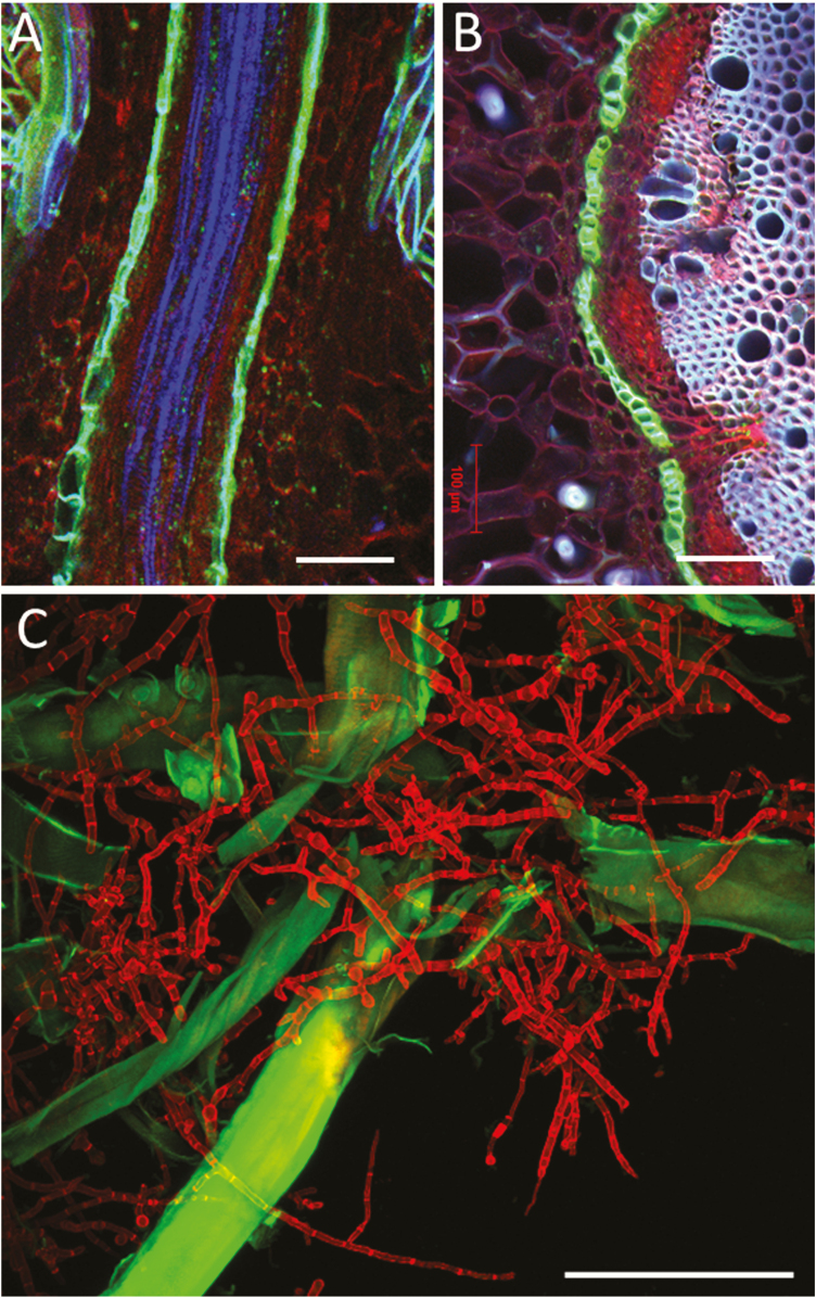
Figure 6.
Multichannel imaging by CLSM. Hand-cut longitudinal (A) and transverse (B) sections of a root of Rhizophora apiculata visualized after double staining with fluorol yellow/Congo red. Single optical sections by CLSM (Zeiss LSM 880, Axio Imager 2, Objective lens: Plan-Apochromat 10/0.45 M27). Merged three channels (ex/em of 405/BP 415–498; 488/BP 543–579; 561/BP 590–630) showing blue for lignin, green for suberized cell walls and red staining of polysaccharides. (C) Maximum image projection of Tricoderma reesei hypha (false colour red) with oxidized lignin-bearing softwood fibres (green). Red hypha (calcofluor) collected with ex/em 405/424–502, green emission from FM 5-95 (fibre) ex/em 488/499–591 (Zeiss LSM 710). The FM 5-95 dye adsorbed on oxidized lignin producing much stronger signal than in membranes. Lignin autofluorescence has insignificant contribution. Bars = 100 µm.

