
Figure 4.
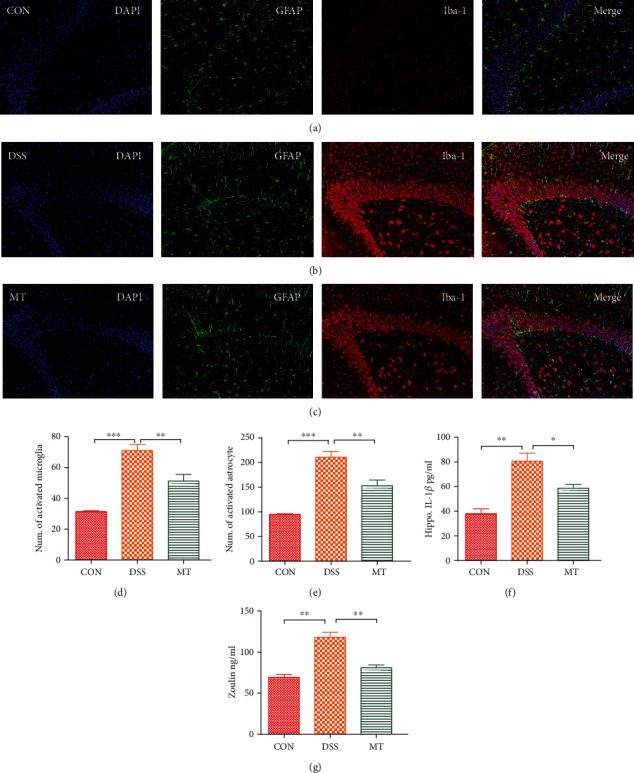
Melatonin-induced attenuation of hippocampal neuroinflammation in DSS rats. (a–c) Micrographs depict labeling of GFAP (green) and Iba-1 (red) in rat hippocampal slices. Nuclear staining was performed with DAPI (blue). (d) The number of cells expressing GFAP, a marker of astrocyte activation. (e) The number of cells expressing Iba1, a marker of microglia activation. (f) Hippocampal IL-1β levels of rats measured with ELISA. (g) Hippocampal zonulin levels of rats measured with ELISA. Data represent the mean ± SEM. ∗p < 0.05, ∗∗p < 0.01, and ∗∗∗p < 0.001. CON = 7; DSS = 5; MT = 6.