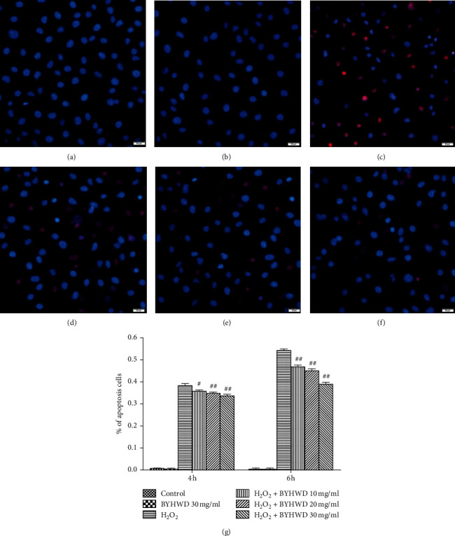
Figure 6.

Effects of BYHWD on H2O2-induced cell apoptosis in HUVECs. Cells were double-stained with cell membrane-permeable (Hoechst 33342; blue) and impermeable (PI; red) DNA labeling fluorochromes. (a) Vehicle medium (control) for 6 h. (b) Preincubation with BYHWD (30 mg/ml) for 6 h. (c) H2O2 (400 μM) for 6 h. (d) Preincubation with BYHWD (10 mg/ml) for 6 h followed by H2O2 for 6 h. (e) Preincubation with BYHWD (20 mg/ml) for 6 h followed by H2O2 for 6 h. (f) Preincubation with BYHWD (30 mg/ml) for 6 h followed by H2O2 for 6 h. (g) The percentage of apoptotic cells for each group and time point. #P < 0.05 and ##P < 0.01 versus the H2O2 group at the same time. Scale bars, 50 μm.
