
FIGURE 4.
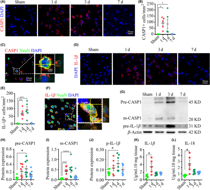
Pro‐inflammatory caspase and cytokines are activated following ischemia. (A‐B) Images and analysis depicted the expression of caspase‐1, n = five mice per group. (C) Images of double‐stained caspase‐1 and NeuN. (D‐E) Series of IF images displayed Il‐1β expression. (F) Images of double‐stained IL‐1β and NeuN. (G‐J) WB images and analysis showed the expression of caspase‐1 and IL‐1β. (K‐L) ELISA analysis of IL‐1β and IL‐18 in the ipsilateral cortex, n = five mice/group. *P < .05, **P < .01, ***P < .001, and ****P = .0001, compared with the sham‐operated group