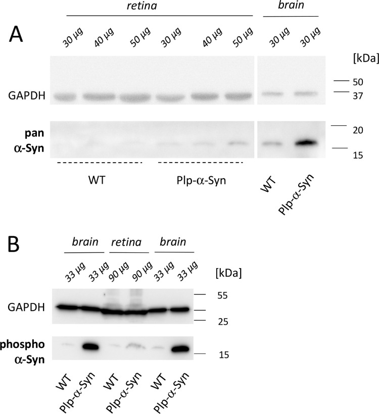
Figure 1.
α-Syn in mouse retinas. α-Syn expression was higher in Plp-α-Syn as compared to wild-type (WT) samples. (A) Retina of both WT and Plp-α-Syn mice compared to brain positive controls. The antibody (pan α-Syn; see Table 1) detected the murine and human forms of α-Syn. (B) The presence of the phosphorylated α-Syn (S129) in the retina, with glyceraldehyde 3-phosphate dehydrogenase (GAPDH) as loading control. In all experiments, the indicated amounts of retinal and brain tissue were loaded. The age of animals was 8 to 10 weeks. The figure shows representative examples of three experiments.

