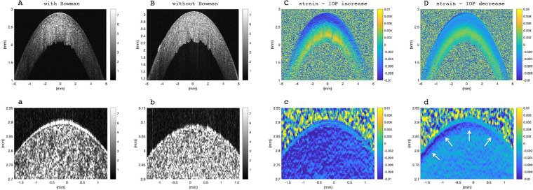
Figure 1.
Corneal tissue at normal IOP (17 mm Hg). Morphological image: (A) with Bowman's layer, (B) without Bowman's layer, removed by excimer laser ablation. Strain distribution with Bowman's layer: (C) resulting from pressure increase by 1 mm Hg, (D) resulting from pressure decrease by 1 mm Hg. Panels (a–d) show a zoomed-in view of the anterior cornea from the image above. White arrows indicate an anterior structure that behaves different than the remaining stroma, particularly during pressure decrease. Corneas with and without Bowman's layer were indistinguishable from strain images.

