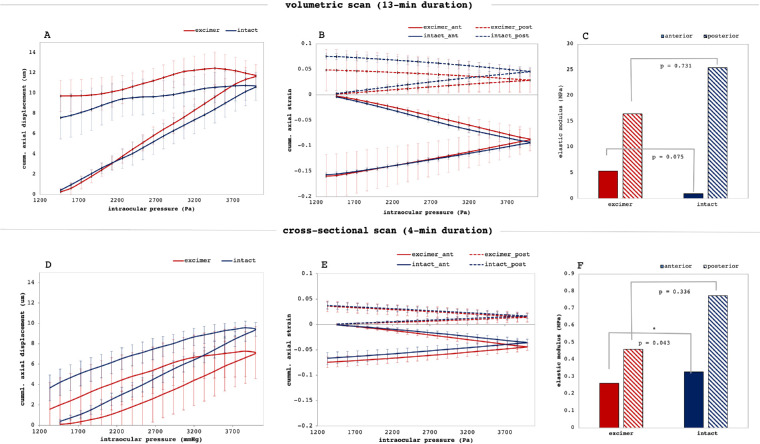
Figure 5.
Biomechanical interpretation. (A,D) Accumulated vertical axial displacement of the central cornea as a function of IOP. Corneas did not fully recover after unloading. (B,E) Accumulated stress-strain diagram in the anterior and posterior cornea as a function of IOP. (C,F) Corresponding elastic moduli of the anterior and posterior cornea (*p<0.05). Error bars are omitted for clarity. Corneal strain and deformation were significantly lower the faster the measurement was conducted corresponding to a higher elastic modulus.

