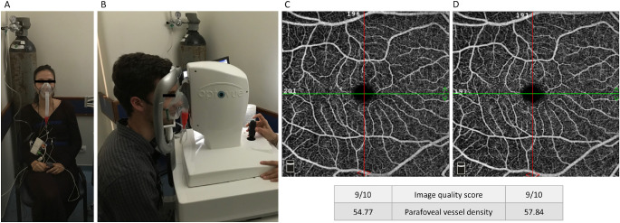
Figure 1.
Exemplar of the setup during the hypoxia challenge test and OCT-angiography examination (A, B) and macular en-face 6- × 6-mm angiograms obtained in baseline conditions (C) and during the hypoxic test (D). The angiograms belong to the healthy volunteer depicted in B. Vessel density increased in hypoxic conditions as expected. Image quality score and parafoveal vessel density are provided according to the built-in angioanalytics software, as described in the Methods section.

