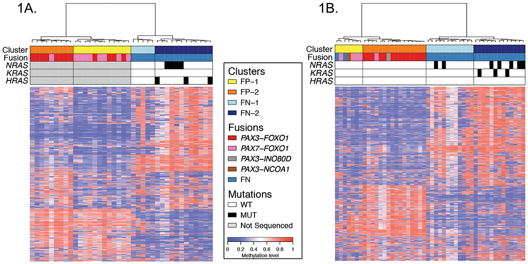
Figure 1. DNA methylation profiling in discovery and validation cohorts identifies molecular subsets in FP and FN RMS.

Heat maps for discovery (A) and validation (B) cohorts displaying the subsets of FP and FN RMS defined by DNA methylation. These displays are based on the top 1% most varied DNA probes across RMS tumors. Differences in the top 1% most varied DNA probes between the discovery and validation cohorts may contribute to the small differences in methylation patterns displayed for the RMS subsets. Fusion status and RAS mutation status are shown in the upper panel in addition to the methylation-defined subsets. Abbreviations: FN, fusion-negative; FP, fusion-positive; WT, wild-type; MUT, mutant-type.
