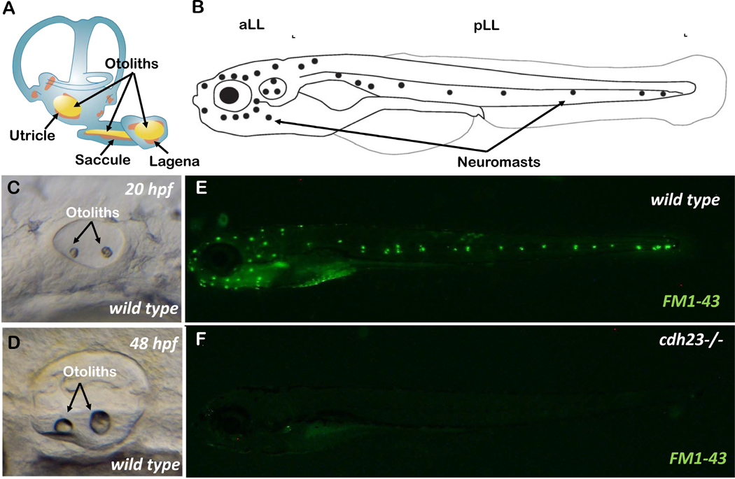
Figure 3.
A) Schematic showing the inner ear of adult zebrafish with semi-circular canal, three sensory epithelia and otoliths. B) Schematic showing the anterior (aLL) and posterior (pLL) lateral lines of a larval zebrafish. Neuromasts are shown by dots. C & D) The maturing inner ear during embryonic development at 20 hours post fertilization (hpf) and 48 hpf, respectively. E & F) Larvae at 5-day post fertilization, stained with FM1–43 live dye, in wild-type and cdh23 mutant animals. Absence of staining in cdh23 mutants is due to dysfunctional mechanotransduction channels.

