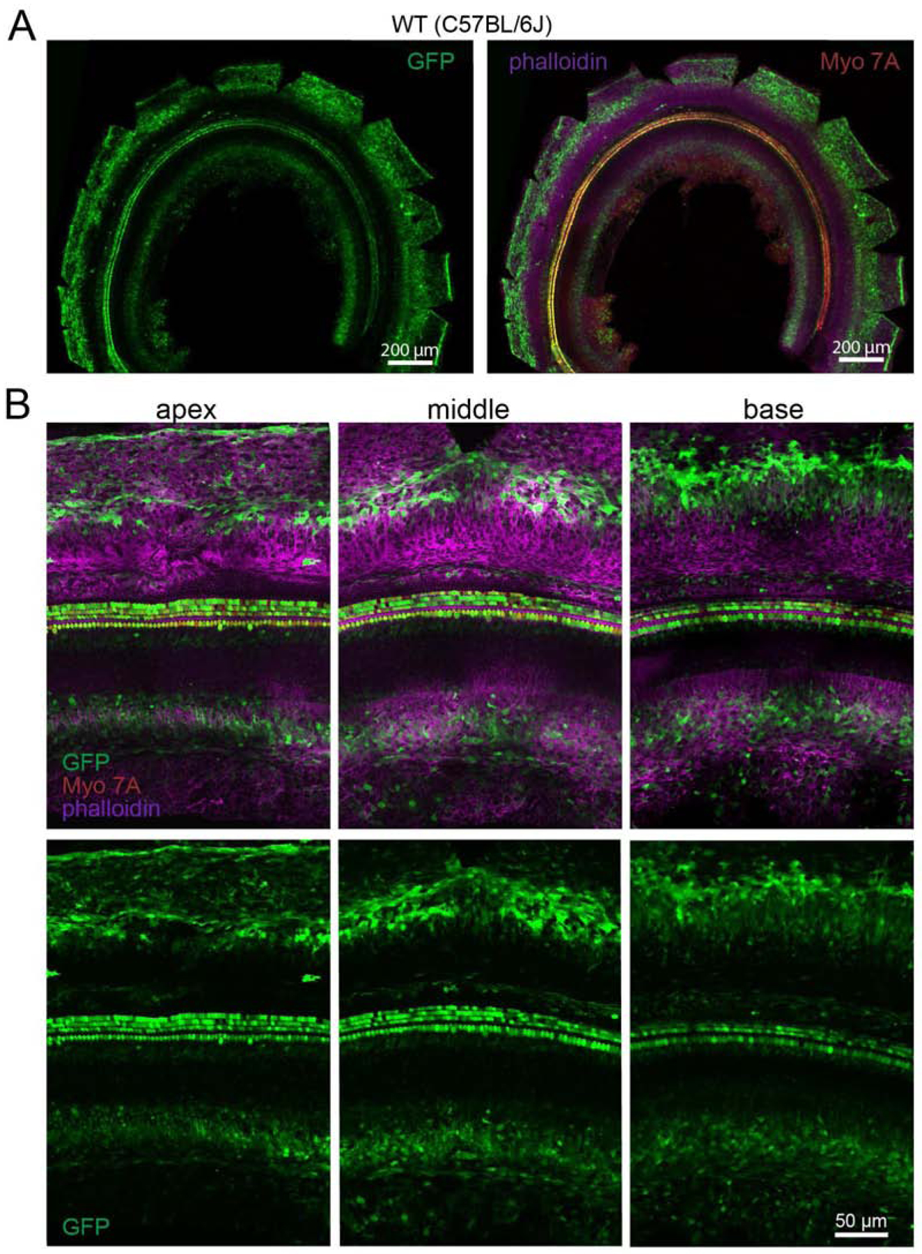
Fig. 3.

Transduction efficiency in C57BL/6J mice of ssAAV9-PHP.B-CBA-GFP after neonatal RWM injection. A) Low-magnification images of the cochlea showing transduction level. Animals were injected at P1 with 5×10¹⁰ VG and the cochlea was dissected and mounted at P6. The left panel shows GFP expression only (green); the right panel shows GFP expression (green), an anti-MYO7A antibody (red) and phalloidin-stained actin (magenta). B) High-magnification images of apical, middle and basal regions of the cochlea. Upper panel shows GFP expression (green), as well as MYO7A (red) and phalloidin (magenta); lower panel shows GFP expression only (green).
