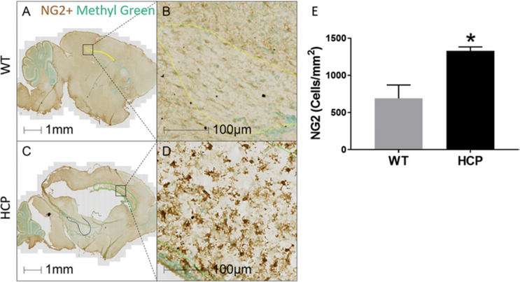
FIG. 5.
Increased NG2 cells in the corpus callosum of hydrocephalic animals as compared to WT animals. Sagittal sections of PND10 WT (A and B) and hydrocephalic (C and D) mice stained with NG2 (brown) and methyl green (green) for nuclei. NG2 density in the corpus callosum significantly increased in animals with hydrocephalus (E). Data are presented as mean ± SEM (n = 3 in each group). *p < 0.05 vs WT.

