
Figure 6: In vivo expression of DAAs/TAAs SOD2, S100A9, and Serpin B1.
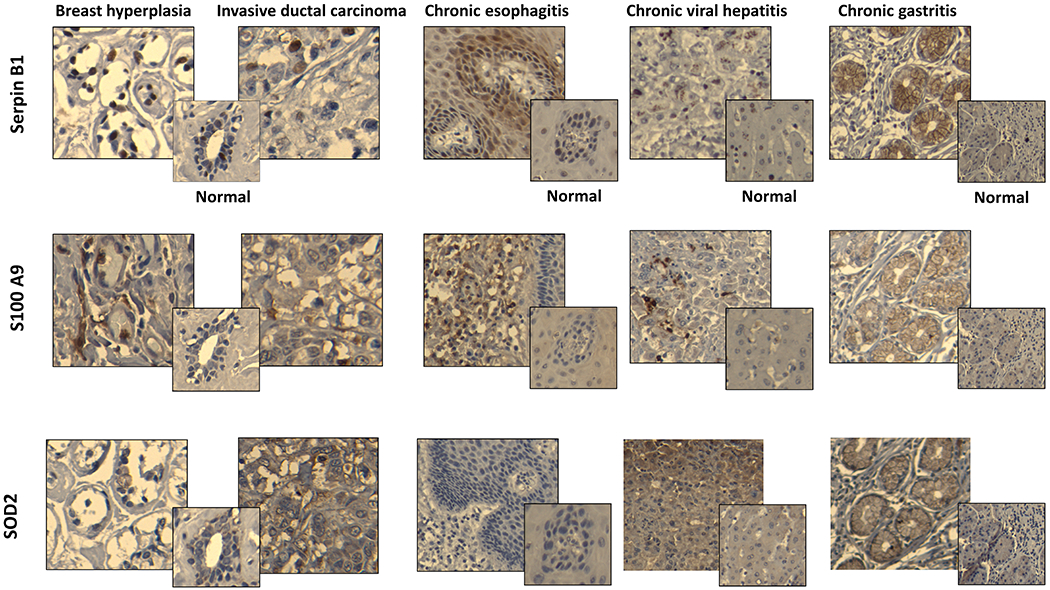
Representative images of paraffin-embedeed tissue sections of breast hyperplasia, breast invasive ductal carcinoma, chronic esophagitis, chronic viral hepatitis, and chronic gastritis stained with relevant antibodies (see Materials and Methods). Corresponding normal tissues stained with the same antibodies are shown in smaller squares. All slides were scanned at 10X magnification in order to select for a high resolution images at 20X.