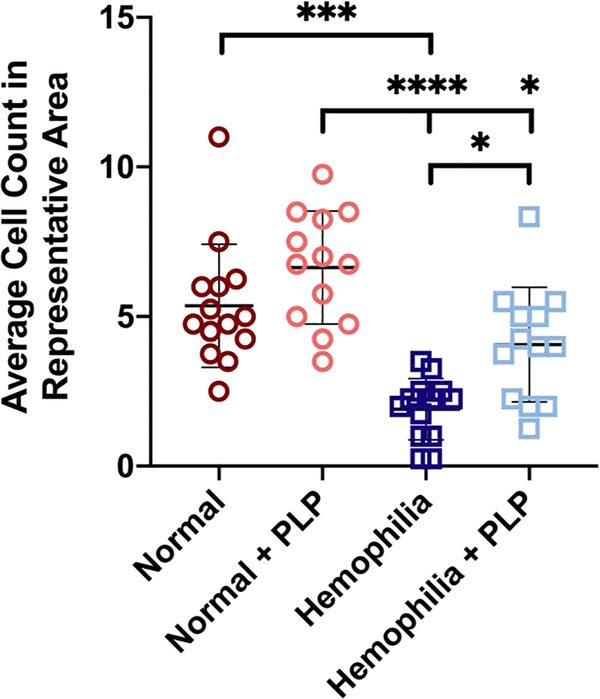
Figure 8: In vitro assessment of fibroblast migration within recalcified platelet rich plasma (PRP) clots.
Fibroblasts were seeded into a collagen/fibrinogen matrix containing tissue factor, which then undergoes an “injury” via a biopsy punch. The injured area is filled with a clot formed from recalcified PRP, and fibroblast migration into the wounded area is observed over time. Average fibroblast counts within representative 300 px2 areas of images obtained 3 days after initial injury. Hemophilia-like structurally deficient PRP clots were able to support similar levels of fibroblast migration as normal clots after incorporation of PLPs and significantly greater fibroblast migration than control hemophilia-like PRP clots. Three independent experiments were conducted; n = 4–5 per group per experiment. Individual data points and mean ± standard deviation are represented for each group. *p < 0.05; ***p < 0.001; ****p < 0.0001.

