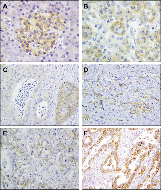
Figure 1.
Expression of IGF1R in normal pancreas and invasive pancreatic ductal adenocarcinoma A. Membranous IGF1R expression occurs in a patchy distribution within pancreatic islets. B. Pancreatic exocrine cells exhibit faint granular expression within the cytoplasm with only rare and weak membranous IGF1R expression. C. Complete absence of IGF1R expression (intensity 0 in 100% of cells, H-score 0) in PDAC with retained expression in an adjacent non-neoplastic islet. D. Patchy, weak (intensity 1 in 50% of cells, H-score 50) IGF1R expression in PDAC. E. Patchy, moderate (intensity 2 in 50% of cells, H-score 100) IGF1R expression in PDAC. F. Diffuse, strong (intensity 3 in 100% of cells, H-score 300) IGF1R expression in PDAC.

