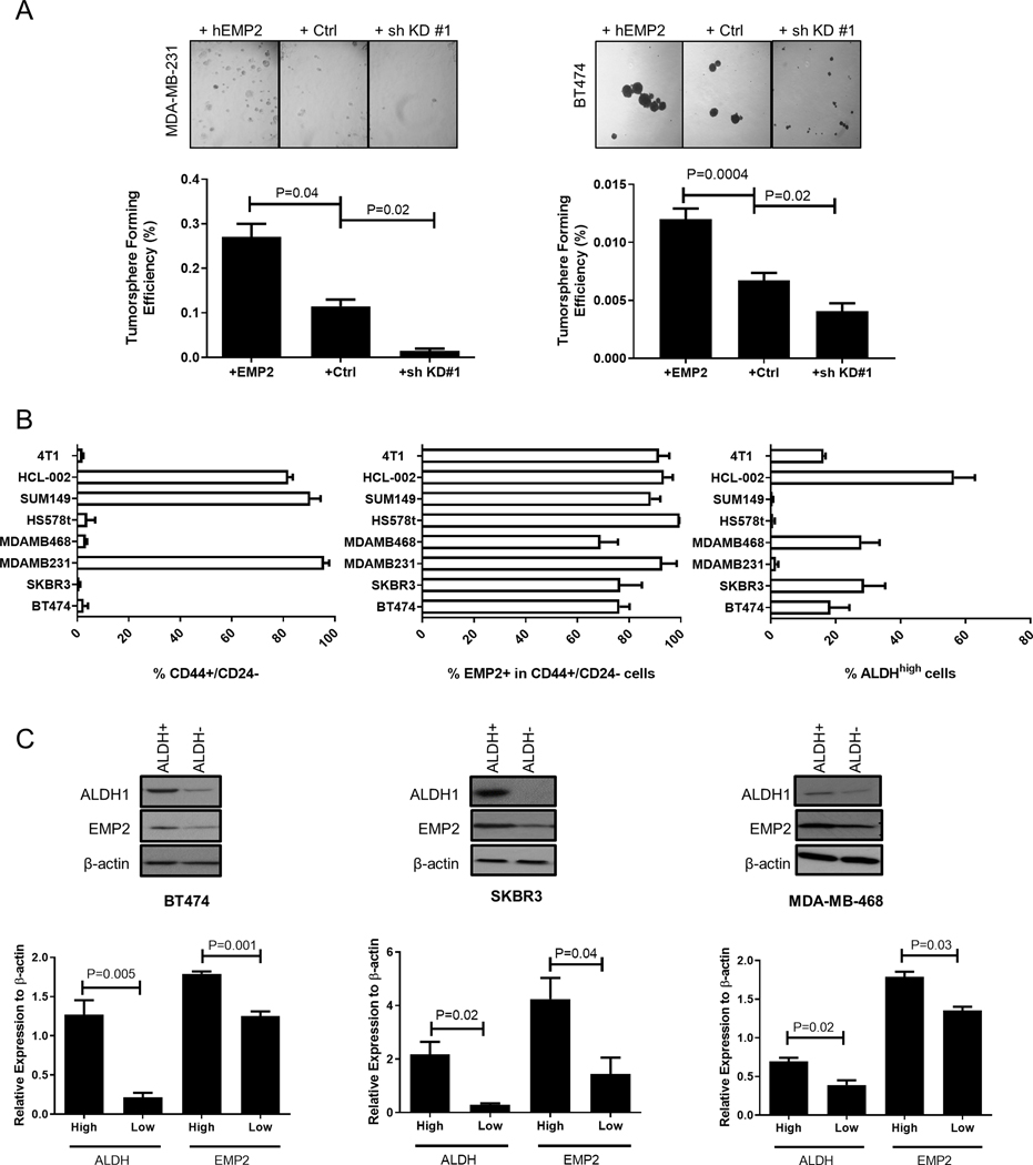
Figure 2. EMP2 expression promotes tumorsphere formation and markers associated with stemness.

A. The effects of EMP2 expression on mammosphere formation was evaluated in MDA-MB-231 and BT474. EMP2 levels directly correlated with increased mammosphere formation. N=4; significance was determined using Student’s t test. B. Breast cancer cells express varying levels of CD44+/CD24- or ALDH activity as determined by the ALDEFLUOR assay. Left, The percentage of CD44+/CD24- cells observed in a panel of breast cancer cell lines as determined by flow cytometry. Middle Panel, Percentage of EMP2+ cells was determined in cells gated on CD44+/CD24- using flow cytometry. Right, Assessment of ALDH activity in a panel of breast cancer cell lines as measured by the ALDEFLUOR assay. N=3 with results expressed as the mean± SEM. C. High levels of ALDH1 correlated with high levels of EMP2 expression. Cells were sorted into ALDHhigh or ALDHlow as determined using the ALDEFLUOR assay and subsequently analyzed for EMP2 and ALDH1 expression by western blot analysis. Results were normalized to β-Actin levels and tabulated as the mean± SEM, with significance determined using Student’s t test. N=3.
