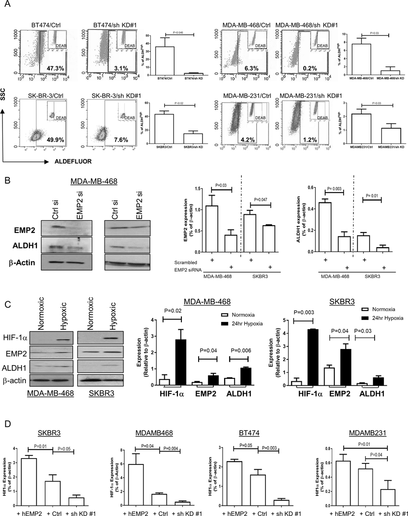
Figure 3. ALDH activity changes relative to EMP2 expression.

A. As a functional readout of ALDH activity, breast cancer cells with endogenous or shRNA knockdown of EMP2 were tested using the ALDEFLUOR assay. A specific inhibitor of ALDH, diethylaminobenzaldehyde or DEAB, was used as a negative control. Higher EMP2 levels correlated with an increase in the number of ALDHhigh cells. B. To independently confirm the regulation of ALDH1 by EMP2, cells were transiently transfected with an EMP2 or scrambled siRNA. Knockdown of EMP2 levels significantly reduced ALDH1 expression in two representative cell lines. N=3, with results tabulated as the mean ± SEM. C. Hypoxia increases ALDH1 and EMP2 levels. MDA-MB-468 and SKBR3 were plated under both normoxic and 0.1% O2 hypoxic conditions. After 24 hours, ALDH1 and EMP2 levels were measured relative to β-Actin. A representative western blot is shown on the left, and results tabulated from three independent experiments shown on the right. Data is expressed as the mean ± SEM. D. SKBR3, MDA-MB-468, BT474 and MDA-MB-231 cells were placed in 0.1% O2 hypoxic chamber. HIF-1α expression was monitored in response to changes in EMP2. All experiments were repeated 3 times, with results expressed as the mean ± SEM. P values show significant differences between groups using Student’s t test.
