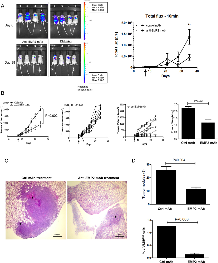
Figure 6. Anti-EMP2 therapy reduces tumor growth and metastasis.

A. In order to create a model of metastasis, MDA-MB-231/Luc+ cells were injected into the left ventricle of Balb/c nude mice, and tumor load was determined using bioiluminescence. Mice were treated systemically, beginning at day 5, with 10mg/kg anti-EMP2 IgG1 or control human IgG. Data was analyzed using the maximum photon flux emission (photons/second) throughout the whole animal. N=4. *, p<0.05 as determined by Student’s t test. B. 5×10⁴ 4T1-FLUC cells were implanted into the mammary fat pad of Balb/c immunocompetent mice and treated twice a week with 10mg/kg control human mAbs or anti-EMP2 mAb starting on day 7 when tumors approached 75–100mm3. The arrow demarks treatment initiation, with grouped and individual data shown. N=8. Two way ANOVA, p=0.002. *, significant by Bonferroni post test. Right, Animals were sacrificed at day 24, and tumors weighed. Mean tumor weight for the control mAb treated group was 1.5±0.3 while the mean tumor weight for the anti-EMP2 mAb was 0.95± 0.1 (p=0.04, Mann Whitney test). N=8. C. In order to assess pulmonary metastases, lungs were removed and stained with hematoxylin and eosin. Representative images are shown. Magnification=400X. D. Top, Enumeration of tumor nodules per 4x field. p=0.004, Student’s t test. Below, For half of the animals, ALDH activity was measured using the ALDEFLUOR assay in the remaining primary tumor. Average results are shown from 4 animals ± SEM, with significance determined using Student’s t test.
