
Figure 11.
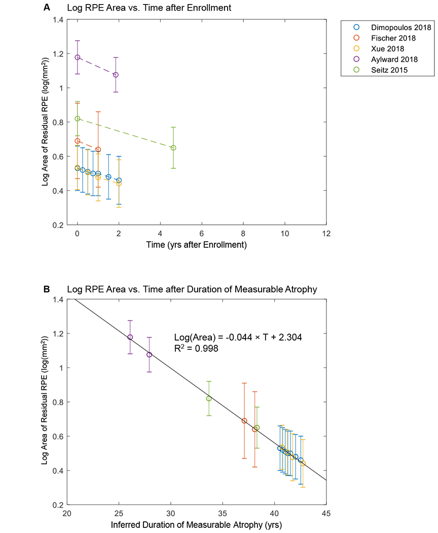
Size of residual retinal pigment epithelium (RPE) as a function of time using aggregate data. A, Aggregate raw data from prior publications (error bars = standard errors). The plot shows a series of lines for the decline of log area of residual RPE in untreated eyes with Choroideremia (CHM). Note that the initial size of residual RPE (time = 0) varies across different studies. B, Data generated by adding a horizontal translation factor (expressed in years) to each data set in (A) to correct for different entry times of the patients at the time of enrollment. Horizontal axis now represents inferred duration of disease rather than years after enrollment into studies. After the introduction of horizontal translation factors, data sets from all 5 studies now fit along a straight line in the area exponential model (r2 = 0.998).