
Figure 15.
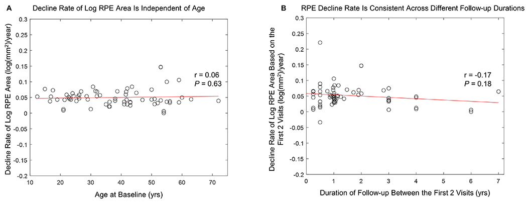
A, Graph showing the decline rate of the log-transformed area of residual retinal pigment epithelium (RPE) was independent of patients’ ages (Pearson’s r = 0.06, P = 0.63). B. Graph showing the decline rate of log-transformed area of residual retinal pigment epithelium (RPE) determined using the first and second visits of each eye was consistent across different durations of follow-up between first and second visits (Pearson’s r = −0.17, P = 0.18). The mean (± SD) RPE decline rate assessed in eyes with ≤1 year follow-up (0.053 ± 0.041 log(mm2)/year) was similar to the decline rate determined in eyes with >1 year follow-up (0.051 ± 0.032 log(mm2)/year) (P = 0.83).