Figure 3.

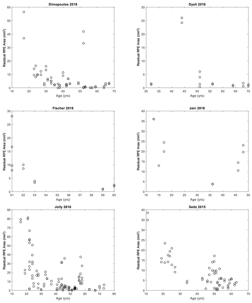
Graphs showing cross-sectional individual participant data extracted from six studies. Each circle represents the area of residual retinal pigment epithelium (RPE) in one eye at the patient’s age of examination.

Graphs showing cross-sectional individual participant data extracted from six studies. Each circle represents the area of residual retinal pigment epithelium (RPE) in one eye at the patient’s age of examination.