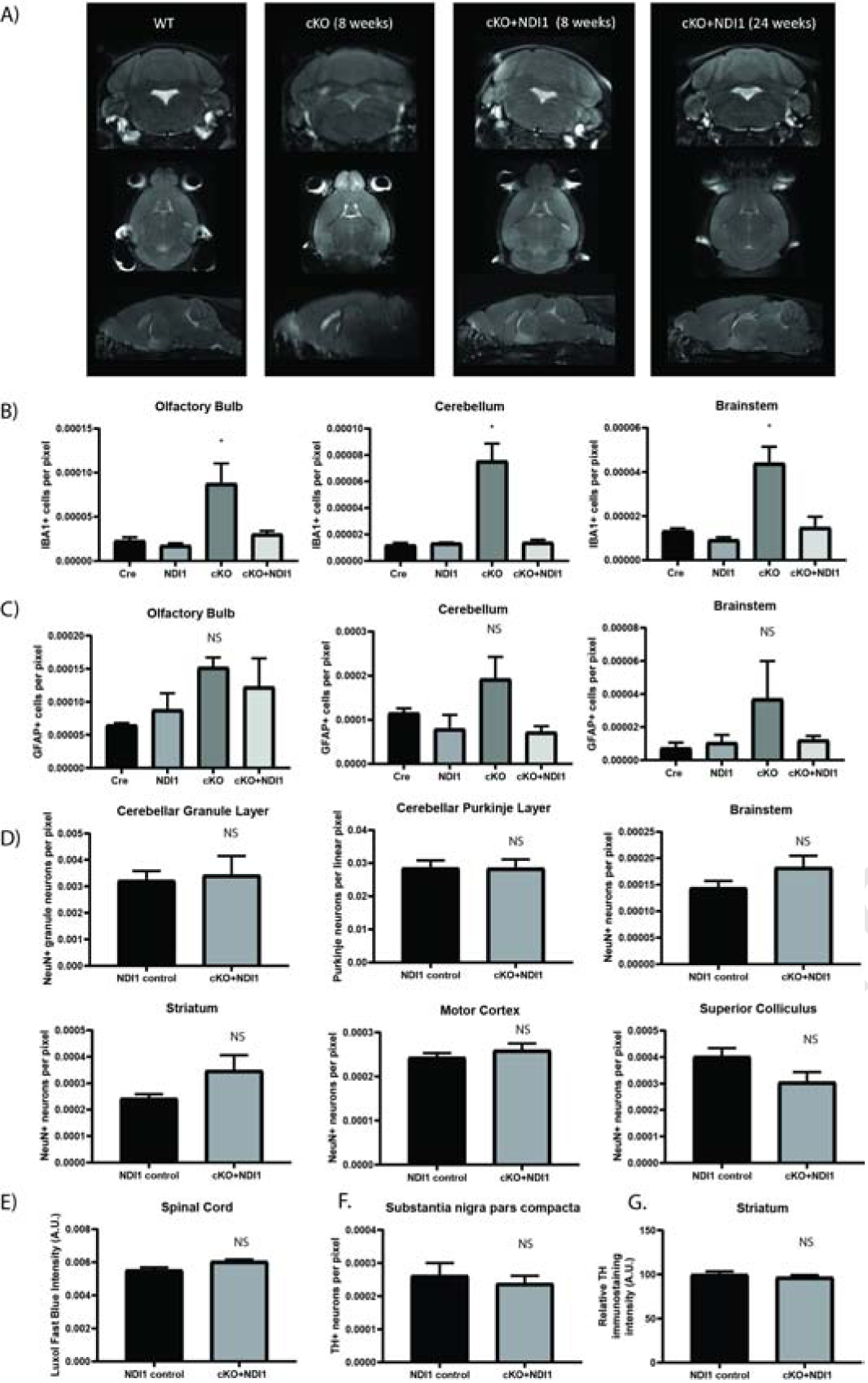
Figure 3.

NDI1 expression prevents the formation of inflammatory glial activation and MRI lesions and does not cause neurodegeneration in a mouse model of Leigh Sydrome. (A) Representative MRI T2-weighted turbo spin echo sequence images. Coronal, horizontal, and sagittal planes are shown at the approximate level where cKO mice exhibit bilateral hyperintense lesions in the olfactory bulb, brainstem, and cerebellum. (B) Quantification of IBA1+ microglia in brain regions demonstrating cKO inflammation at 7–8 weeks of age (N=3–4, ANOVA with Dunnet’s test vs WT P<0.05*). See also Fig. S3. (C) Quantification of GFAP+ astrocytes at 7–8 weeks of age (N=3). (D) Quantification of neurons from motor control brain regions at over 1 year of age (N=6). (E) Quantification of spinal cord Luxol fast blue staining (N=3). (F) Quantification of tyrosine hydroxylase+ dopaminergic neurons (N=6) (G) Tyrosine hydroxylase+ immunoreactivity in striatum (N=5–6). (C-G) ANOVA and/or T-test not significant. Data represent mean ± SEM. See also Fig. S3.
