FIGURE 2.
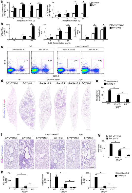
ILC2s do not fully account for chronic lung disease after viral infection. (a) Levels of total cells and ILC2s in the lungs of WT mice at indicated times after infection with SeV or Se-UV as determined by flow cytometry. (b) Levels of ILC2 markers (Il13, Gata3, and Rora) mRNA in ILC2s that were FACS-isolated from lung and treated with IL-33 (0-20 ng/ml). (c) Cytograms for ILC2s (Lin−CD90.2+ST2+) in WT and Il7rwt/Cre-Rorafl/fl mice at 49 d after SeV or SeV-UV. (d) Hematoxylin and eosin staining of full-lung sections from indicated mouse strains at 49 d after SeV or SeV-UV. (e) Quantitation of tissue staining in (d) using image-analysis software. (f) PAS and hematoxylin staining of lung sections for conditions in (d). (g) Quantitation of tissue staining in (f). (h) Levels of indicated mRNAs in lung tissue for conditions in (d). All data are representative of three separate experiments (mean and s.e.m.) with at least 5 mice per condition in each experiment. *P<0.01.

