FIGURE 6.
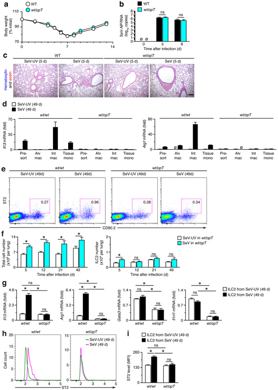
Tissue macrophages regulate ILC2 accumulation and activation in chronic lung disease after viral infection. (a) Body weights at indicated times after infection with SeV in WT and wt/opT mice. (b) Corresponding viral loads in lungs for conditions in (a). (c) Hematoxylin and eosin staining of lung sections for indicated conditions. Scale bar, 500 μm. (d) Levels of Il13 and Arg1 mRNA in FACS-isolated macrophage populations from lungs of wt/wt and wt/opT mice at 49 d after SeV or SeV-UV. (e) Flow cytograms for ILC2s from wt/wt and wt/opT mice at 49 d after SeV or SeV-UV. (f) Quantitation of total cell and ILC2 levels from lungs of wt/opT mice at 5-49 d after SeV or SeV-UV using flow cytometry conditions in (e). (g) Levels of Il13, Gata3, Arg1, and Il1rl1 mRNA in FACS-isolated ILC2s from wt/wt and wt/opT mice at 49 d after SeV or SeV-UV. (h) Flow histograms for ST2 expression in ILC2s from lungs of wt/wt and wt/opT mice at 49 d after SeV infection or SeV-UV. (i) Levels of ST2 based on MFI for conditions in (h). All data are representative of three separate experiments (mean and s.e.m.) with at least 5 mice per condition in each experiment. *P<0.01.

