Figure 3.

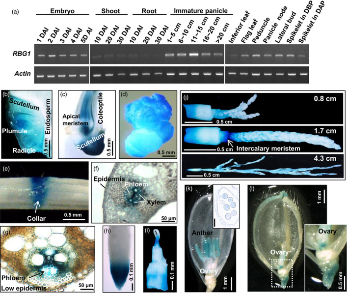
RBG1 is expressed preferentially in rice tissues with cell division activities. (a) Total RNAs were extracted from various tissues at different developmental stages of WT rice, and subjected to RT‐PCR analyses using gene‐specific primers (Table S4). DAI, day after imbibition; DBP, day before pollination; DAP, day after pollination. (b–l) Various tissues from transgenic rice carrying RBG1:GUS were sectioned and stained for GUS activity. (b) Longitudinal section of embryo at 1 DAI. (c) Longitudinal section of shoot at 5 DAI. (d) Cultured callus. (e) Leaf sheath showing collar at 15 DAI. (f) Cross section of leaf sheath showing vascular bundle at 20 DAI. (g) Vascular bundle at 30 DAI. (h) Root tip at 20 DAI. (i) Shoot apical meristem at 20 DAI. (j) Immature panicles at different developmental stages, that is 0.8, 1.7 and 4.3 cm in length, and intercalary meristem of panicle node. (k) Spikelet before pollination. The inset image shows pollen in a cross‐sectioned anther. Scale bar in inset: 50 µm. (l) Spikelet after fertilization. The inset image shows an enlarged ovary. Scale bar in inset: 500 µm.
