Figure 4.

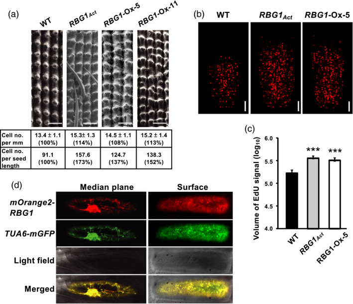
An increase in cell number caused by increased cell division is responsible for the increase in seed size in RBG1 overexpression lines. (a) Examination of abaxial epidermal cells of hull lemma by scanning electron microscopy revealed a significantly greater number of cells per unit length in RBG1 overexpression lines than in WT. Scale bar = 100 μm. (b) Roots of seedlings at 8 DAI were stained with 2 µm EdU and visualized by Z‐stack serial sections using confocal microscopy. Single optical sections of 6 μm (optical depth) on the median plane of rice root tips were captured. Scale bar = 50 μm. (c) Quantification of average EdU signal intensity of the meristem region presented on a logarithmic scale. n = 9, 20 and 18 for WT, RBG1Act and RBG1‐Ox‐5 lines, respectively. (d) Onion epidermal cells were co‐transfected with the constructs Ubi:mOrgange2‐RBG1 and Ubi:TUA6‐eGFP by particle bombardment. Left and right panels show the middle plane and the surface of the same cell, respectively.
