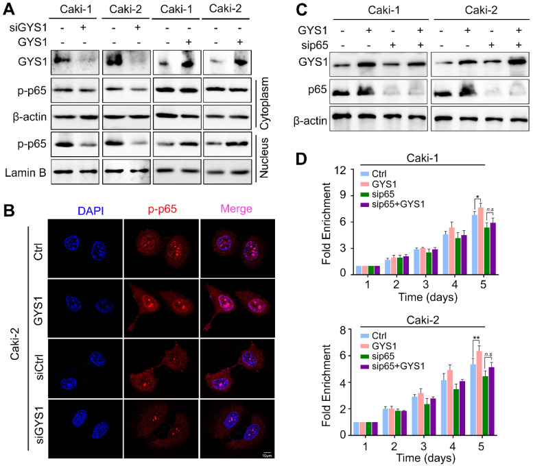
Figure 5.
GYS1 activates the NF-κB signaling pathway. (A) Western blot revealed the expression of p-p65 in the cytoplasm and nucleus. β-Actin and Lamin B were used as internal references in the cytoplasm and nucleus, respectively. (B) IF images indicating the subcellular localization of p-p65 (red). The nuclear outline was stained by DAPI (blue) in Caki-2 cells. (C) Expression of p65 and p-p65 was measured by western blotting following transfection of p65 siRNA in rescue experiments. (D) Cell viability was assessed by CCK8 in rescue experiments. Statistical data are represented as the mean ± SD. *P < 0.05, **P < 0.01.

