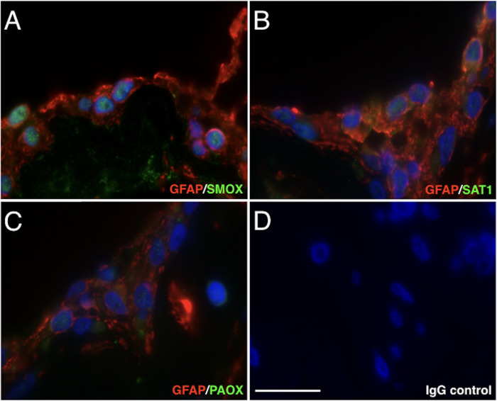
Figure 1.
Immunofluorescence staining of SMOX, SAT1, and PAOX in fibrovascular tissues of patients with PDR. (A) Green, SMOX (Alexa Fluor 488); red, GFAP (Alexa Fluor 546). (B) Green, SAT1; red, GFAP. (C) Green, PAOX; red, GFAP. (D) Negative control (rabbit and mouse normal IgG) in sequential sections. Scale bar = 20 µm.

