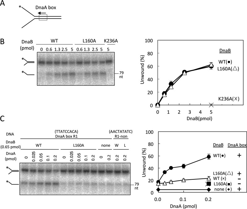
Figure 5.
DnaB helicase activity on fork DNAs. A, structure of fork DNA bearing DnaA box R1; direction of the box sequence (5′-TTATCCACA) is indicated. *, 5′-end 32P label. B, fork DNA (10 fmol, 1.1 nm) with a 32P-labeled 5′-end (*) was incubated at 37 °C for 30 min with DnaB (0–5 pmol or 560 nm; WT, L160A, or K236A). DNA was extracted and analyzed by PAGE. The proportion of ssDNA (as a fraction of total DNA) was plotted. Experiments were performed in duplicate, and a representative gel image and means ± S.D. (error bars) are shown. DnaB K236A, an ATPase-deficient mutant (17), was used as a negative control. C, fork DNAs (10 fmol, 1.1 nm) with DnaA box R1 sequence (R1) or a nonsense sequence (non) were incubated at 37 °C for 30 min with DnaB (0.65 pmol, 72 nm; WT or L160A) and DnaA (0–0.2 pmol or 22 nm). DNA was analyzed as above. DnaA box R1 and nonsense sequences are TTATCCACA and AACTATATC, respectively. W, WT; L, L160A.

