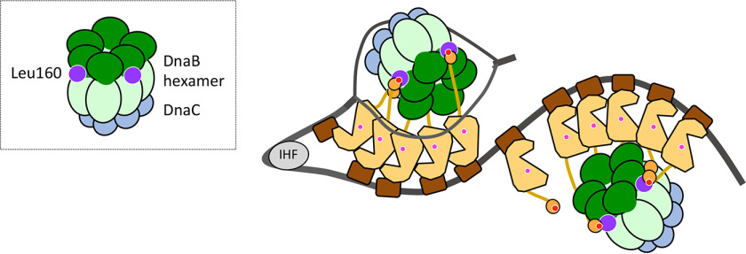
Figure 8.
Model of DnaB loading via interaction between DnaB Leu160 and DnaA Phe46. A model of the structure of the open complex is shown as in Fig. 1. In addition, DnaB (NTD (dark green) and LH-CTD (pale green)), DnaC (cyan), DnaB Leu160 site (purple), and DnaA Phe46 site (dark red) are shown. Two or three protomers of the DnaB hexamer bind to domain I in DnaA pentamers on oriC, which recruits two DnaB hexamers and tethers them in a particular orientation. This conformation underlies the functional interaction of DnaB–DnaC complexes with the DnaA domain III site containing His136 and the unwound region of oriC, promoting DnaB loading onto the oriC region.

