Figure 2.

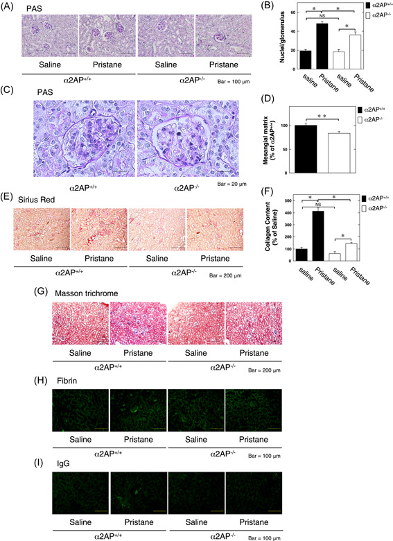
α2AP deficiency attenuated the development of lupus nephritis in the pristane‐induced lupus mouse model. A, Represetative kidney sections from saline or pristane‐treated α2AP+/+ and α2AP−/− mice (periodic acid Schiff [PAS] stain). B, The number of nuclei per glomerulus in the saline or pristane‐treated α2AP+/+ and α2AP−/− mice (n = 10). C, The magnified image of kidney sections from pristane‐treated α2AP+/+ and α2AP−/− mice (PAS stain). D, Mesangial matrix was measured by PAS stain in the kidney from pristane‐treated α2AP+/+ and α2AP−/− mice (n = 4). Mesangial matrix area was expressed as a percent of the observed with control in the pristane‐treated α2AP+/+ mice. E, Representative kidney sections from the saline or pristane‐treated α2AP+/+ and α2AP−/− mice (sirius red stain). F, The collagen content in the kidney from saline or pristane‐treated α2AP+/+ and α2AP−/− mice (n = 4). G, Represetative kidney sections from the saline or pristane‐treated α2AP+/+ and α2AP−/− mice (masson trichrome stain). H, Paraffin sections of the kidney of the α2AP+/+ and α2AP−/− mice were stained with anti‐fibrin antibodies. I, Paraffin sections of the kidney of the α2AP+/+ and α2AP−/− mice were stained with immunoglobulin G antibodies. The data represent the mean ± SEM. *P < .01. **P < .05. α2AP, alpha2‐antiplasmin; NS, not significant
