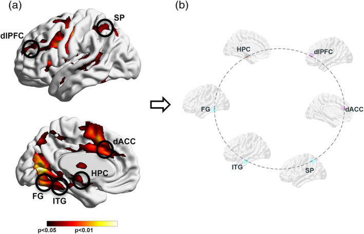
FIGURE 1.

(a) The results of a conjunction analysis (SCZ ∩ HC) are projected to bilateral lateral and medial cortical surfaces. The significance peaks (insets) constitute a common substrate of activation across groups and conditions. These were harvested for subsequent dFC analyses, to avoid connectivity estimates from being confounded by activation differences, and to base dFC estimates on statistically filtered fMRI data. The harvested peaks represented the dorsolateral prefrontal cortex (dlPFC), the dorsal anterior cingulate (dACC), the hippocampus (HPC), the superior parietal cortex (SPC), the fusiform gyrus (FG), and the inferior temporal gyrus (ITG). (b) The schematic connectomic ring provides the framework for subsequent depiction of dFC results (Figures 3, 4, 5). The nodes are color coded by functional clusters; frontal/executive function (dlPFC, dACC; light purple), medial temporal lobe (HPC; gray), and unimodal function (FG, ITG, SP; teal). dFC, directional functional connectivity; HC, healthy controls; SCZ, schizophrenia
