FIGURE 1.
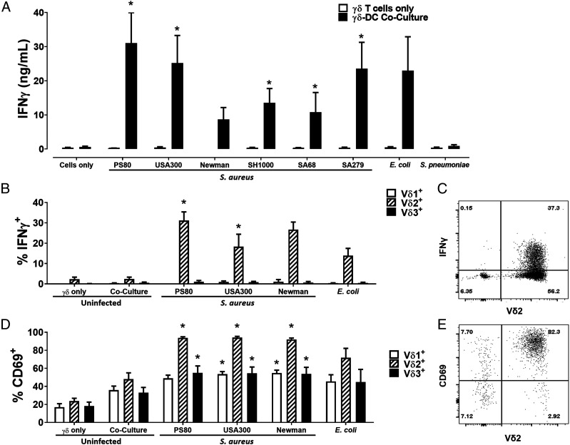
Human blood–derived Vδ2+ cells produce IFN-γ in response to S. aureus–infected DCs. γδ T cells and DCs (5 × 10⁵ cells/ml) were infected with S. aureus (strains PS80, USA300, Newman, SH1000, SA68, and SA279), E. coli (strain CFT073), and S. pneumoniae (strain 6301) at multiplicity of infection 10 for 3 h before elimination of extracellular bacteria by gentamicin treatment. DCs were then cocultured with uninfected γδ T cells (5 × 10⁵/ml) for 24 h. IFN-γ levels in culture supernatants at 20 h were assessed by ELISA (A). Results are expressed as mean IFN-γ concentration in culture supernatants ± SEM. Cells from cocultures with DCs infected with selected S. aureus strains (PS80, USA300, and Newman) and E. coli were treated with BFA for a further 4 h, and IFN-γ expression by Vδ1+, Vδ2+, and Vδ3+ cells was assessed by flow cytometry (B). Results are expressed as mean IFN-γ+ cells within total live singlet cells of each subset ± SEM. Representative FACS plot for PS80 infection is shown (C). CD69 expression on Vδ1+, Vδ2+, and Vδ3+ cells was also assessed in separate experiments (D). Results are expressed as mean CD69+ cells within total live singlet cells of each subset ± SEM. Representative FACS plot for PS80 infection is shown (E). n = 2–3 DC donors per group; n = 7–12 γδ T cell donors per group. Statistical analysis by pairwise Wilcoxon signed-rank test, with all columns compared directly to cells only (A) or uninfected γδ–DC coculture (B and D). *p < 0.05.

