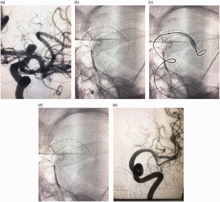
Figure 1.
(a) Left internal carotid digital subtraction angiography anteroposterior projection showing an internal carotid termination aneurysm inclined on the A1 segment. (b) Native image showing inadequately opened proximal end of the flow diverter. (c) Native image showing balloon inflation of the proximal end of the flow diverter. (d) Native image post balloon dilatation showing fully opened proximal end of the pipeline device. (e) Follow-up 12 months digital subtraction angiography anteroposterior projection showing non-filling of the aneurysm as well as the A1 segment.

