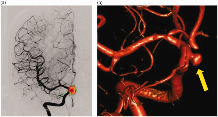
Figure 2.
Example of a true positive result: (a) the 5 mm saccular aneurysm of the right Internal carotid artery, ophthalmic segment, was correctly detected on an anteroposterior view. The user-defined region of interest (ROI; circle with diagonal red lines) and the ROI drawn by the deep learning software (red dashed circle) show a perfect overlap centred over the aneurysm. The subtle green heat map seen in the periphery of the red ROI indicates the saliency map provided by the software. The 3D rotational digital subtraction angiography (DSA) image (b) serves as the gold standard for aneurysm detection (arrow).

