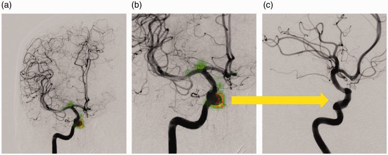
Figure 3.
Example of a false positive result: (a) the deep learning software incorrectly labelled the ophthalmic segment of the internal carotid artery (ICA) as an aneurysm on an anteroposterior view (red dashed circle). The green saliency maps reveal weighting around the ophthalmic segment and at the ICA bifurcation. The result seems plausible given the course of the artery and rounded shape protruding from the vessel which can be appreciated on the enlarged image (b). The additional oblique view (c) at the same level (arrow) confirms the location to be aneurysm-negative.

