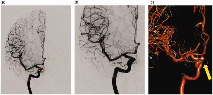
Figure 4.
Example of a false negative result: the aneurysm of the Internal carotid artery (ICA), ophthalmic segment, was not detected by the deep learning software. On image (a) only the user-defined region of interest (ROI; red) can be found. The saliency map (green) indicates weighting near this aspect of the Internal carotid artery, however the structure did not meet the deep learning network’s threshold. Further weighting is seen at the ICA bifurcation. The image without the ROI (b) makes the result seem plausible as the aneurysm appears hidden by the course of the artery on the anteroposterior view. 3D rotational digital subtraction angiography (DSA) (c) image reveals the aneurysm (arrow).

