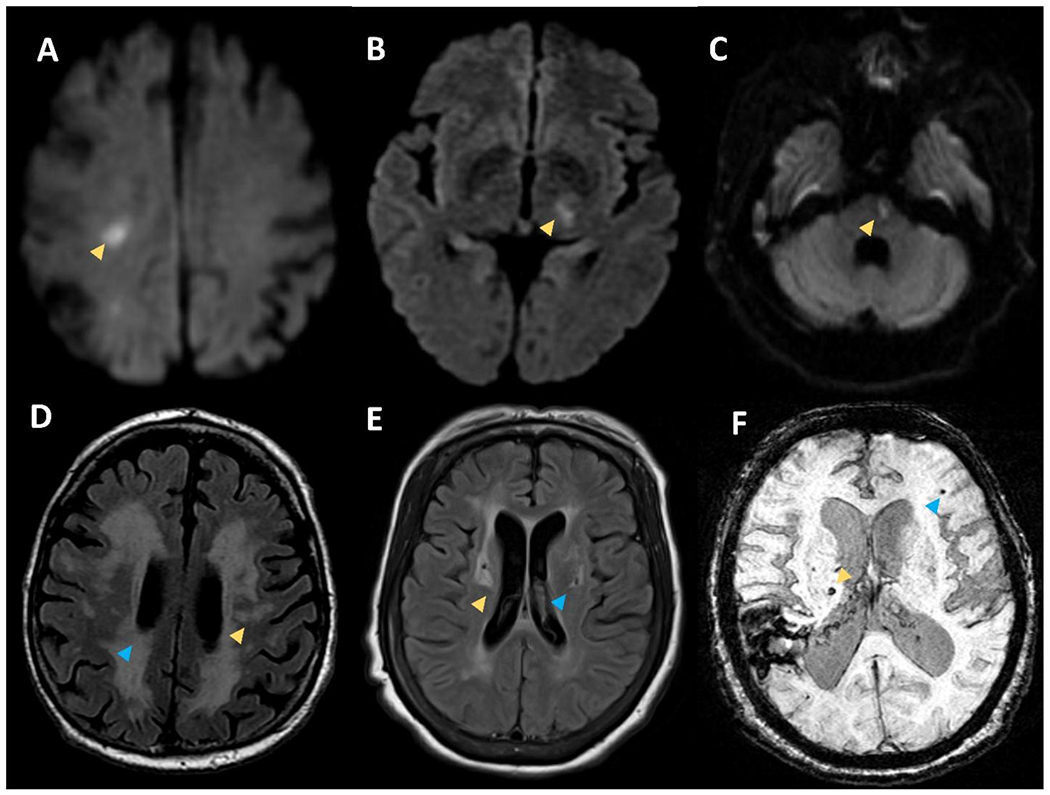
Figure 1.

Small infarcts on DWI sequences within the right subcortical white matter (A), left thalamus (B), and left pons (C) consistent with acute lacunar strokes. Panel D shows T2 FLAIR sequence with white matter hyperintensities in the subcortical white matte (yellow arrow) and periventricular white matter (blue arrow). Panel E shows T2 FLAIR sequence with white matter hyperintensities on in the basal ganglia (yellow arrow) and deep chronic lacune with a rim of hyperintensity (blue arrow). Panel F shows susceptibility weighted imaging with superficial microbleed (blue arrow) and deep microbleed (yellow arrow).
