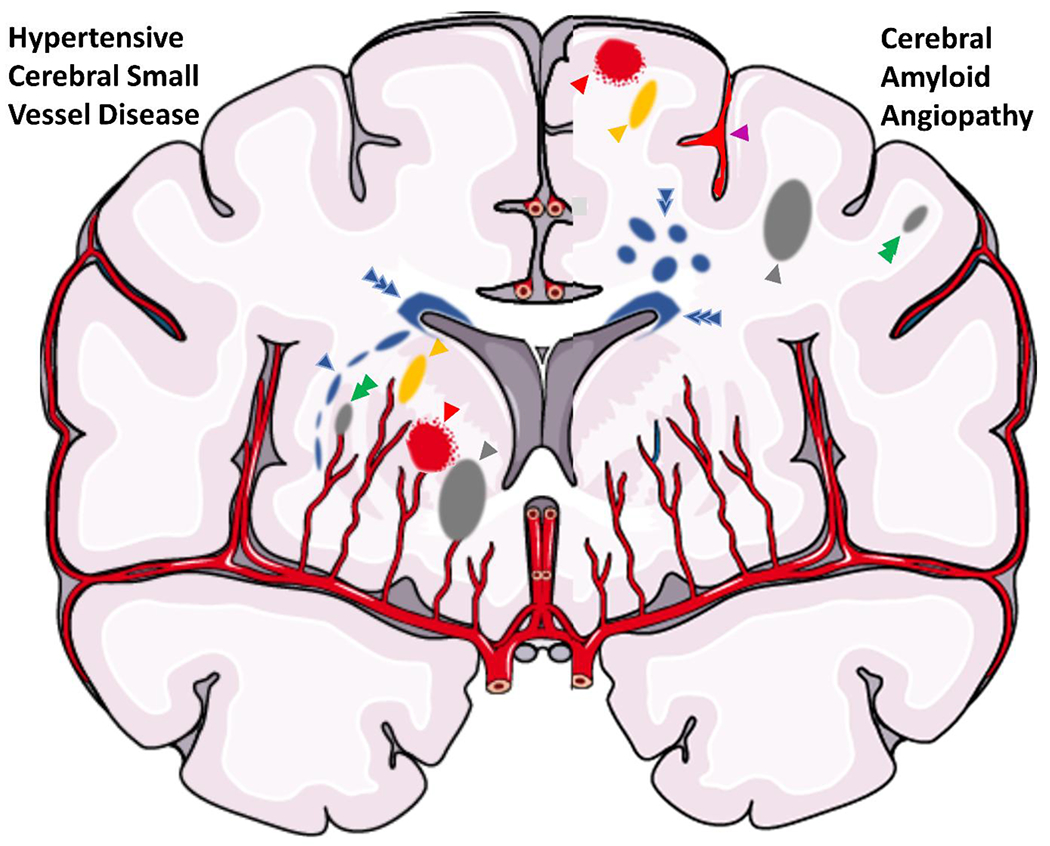
Figure 2.

Illustration of a coronal brain section with parenchymal findings of cerebral small vessel disease. The right hemisphere includes findings suggestive of hypertension-related small vessel disease, including a peri-basal ganglia white matter hyperintensity (blue arrow), dilated perivascular space (yellow arrow), microinfarct (green arrow), deep lacune (gray arrow), and microbleed (red arrow). The left hemisphere includes findings suggestive of cerebral amyloid angiopathy, including a microbleed (red arrow), sulcal siderosis (purple arrow), dilated perivascular space (yellow arrow), subcortical white matter hyperintensity spots (double blue arrow), lobar lacune (gray arrow), and microinfarct (green arrow). There is also perivascular white matter hyperintensity (triple blue arrow), which is associated with both small vessel diseases.
