
Figure 1. DNA methylation sustains tumor-induced MDSC accumulation in vivo.
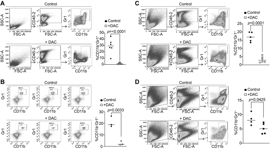
A. AT3 tumor cells (2.5x10⁵ cells/mouse) were injected into the mammary gland of C57BL/6 mice. Tumor-bearing mice were treated with saline (control, n=6) and DAC (2 mg/kg body weight, n=7) by i.v. injection at days 21-23 after tumor cell injection once every 2 days for 2 times. Mice were sacrificed 1 day after the last treatment. Spleens cells were stained with CD45.2−, CD11b−, and Gr1-specific antibodies. All CD45.2+ cells were gated and analyzed for CD11b+Gr1+ cells. Shown are gating strategy (left panel) and quantification of CD11b+Gr1+ cells (right panel). B. AT3 tumor-bearing mice were treated with saline (control, n=3) and DAC (2 mg/kg body weight, n=3) as in A. The peripheral blood was collected and processed for white blood cells. The cells were then stained and analyzed as in A. C. AT3 tumor-bearing mice were treated with saline (control, n=5) and DAC (2 mg/kg body weight, n=6) as in A. Tumor tissues were collected, digested with collagenase to make single cells. The cells were then stained with CD45.2−, CD11b−, and Gr1-specific antibodies. All CD45.2+ cells were gated and analyzed for CD11b+Gr1+ cells. Shown are gating strategy (left panel) and quantification of CD11b+Gr1+ cells (right panel). D. CT26 cells (2x10⁵) were injected s.c. to BALB/c mice. The tumor-bearing mice were treated with saline (control, n=4) and DAC (2 mg/kg body weight, n=5) at days 21 and 23 after tumor cell injection. Mice were sacrificed at day 24. Tumor tissues were collected, digested with collagenase to make single cells. The cells were then stained with CD45.2−, CD11b−, and Gr1-specific antibodies. All CD45.2+ cells were gated and analyzed for CD11b+Gr1+ cells. Shown are gating strategy (left panel) and quantification of CD11b+Gr1+ cells (right panel).