
Figure 7. Autocrine IL6 activates STAT3 to up-regulates DNMT1 and DNMT3b in MDSCs.
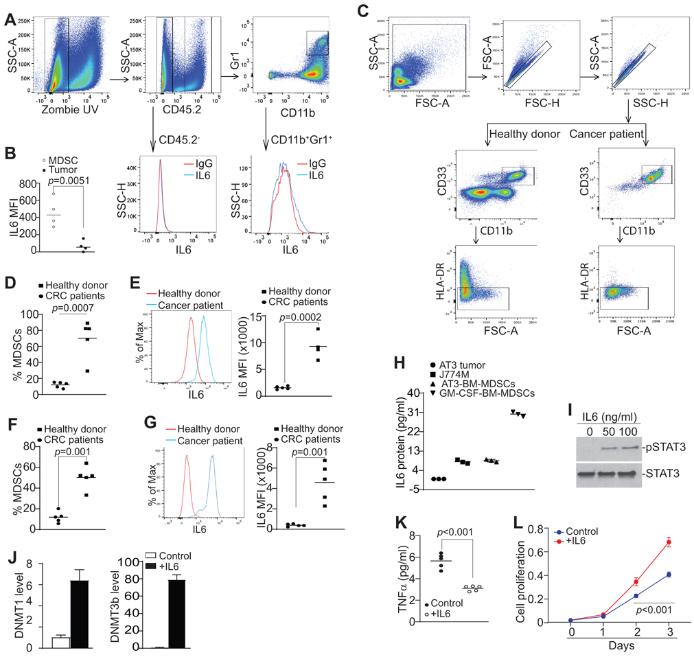
A. CT26 cells were orthotopically transplanted to cecal wall of BALB/c mice (n=4). The tumor tissues were collected, digested in collagenase solution to make single cells. The single cell mixtures were then stained with IgG isotype, CD45.2−, CD11b−, Gr1−, and IL6-specific mAbs plus Zombie UV, and analyzed by flow cytometry. The gating strategy is shown. The CD45.2− tumor cells and CD11b+Gr1+ cells were gated and analyzed for IL6 protein MFI, respectively. Shown are representative results from one of four mice. B. Quantification of IL6 protein level (MFI) in tumor cells and tumor-infiltrating MDSCs. IL6 MFI is calculated as IL6 MFI of the anti-IL6 mAb-stained sample – IgG MFI of the same sample. C. Peripheral blood specimens were collected from healthy donors (n=5) and colorectal cancer patients (n=5). Buffy coat was processed, stained with CD11b−, CD33−, HLA-DR−, and IL6-specific antibodies, and analyzed by flow cytometry. The CD11b+CD33+HLA-DR− cells were gated as shown (top two panels). D. Quantification of CD11b+CD33+HLA-DR− MDSCs as shown in C. Each dot represents % MDSCs from one donor or patient as described in Table S1. E. The representative overlay of IL6 MFI of MDSCs of one healthy donor and one colorectal cancer patient. Quantification of MFIs were shown at the right panel. Each dot represents % MDSCs from one donor or patient as described in Table S1. F & G. Peripheral blood specimens were collected from five more healthy donors (n=5) and colon cancer patients (n=5) as described in Table S2 and repeated the analysis of IL6 in MDSCs as in C-E. H. AT3 tumor cells and J774M cells were cultured for 24h and the culture medium was collected and analyzed for IL6 protein. To induce MDSC from BM cells, BM cells were isolated from C57BL/6 mice and cultured with AT3 tumor cell-conditioned medium or GM-CSF for 5 days. The culture supernatants were then collected and analyzed for IL6 protein level. I. J774M cells were treated with recombinant IL6 protein at the indicated doses for 1.5h and analyzed by Western blotting analysis for the indicated proteins. J. J774M cells were treated with recombinant IL6 protein (100 ng/ml) for 24h and analyzed for DNMT1 and DNMT3b expression by qPCR with Rpl13a as an internal control. K. J774M cells (1x10⁵/well) were seeded in a 96-well plate and cultured with or without 100 ng/mL IL-6 (R&D). Supernatants were collected 24h later and measured for TNFα by ELISA. F. J774M cells (1x10³ cells/well) were seeded in a 96 well plate and treated with 100 ng/mL IL-6. Cell proliferation at indicated time points was measured using AqeousOne reagent according to manufacturer’s instructions (Promega).