
Figure 3:
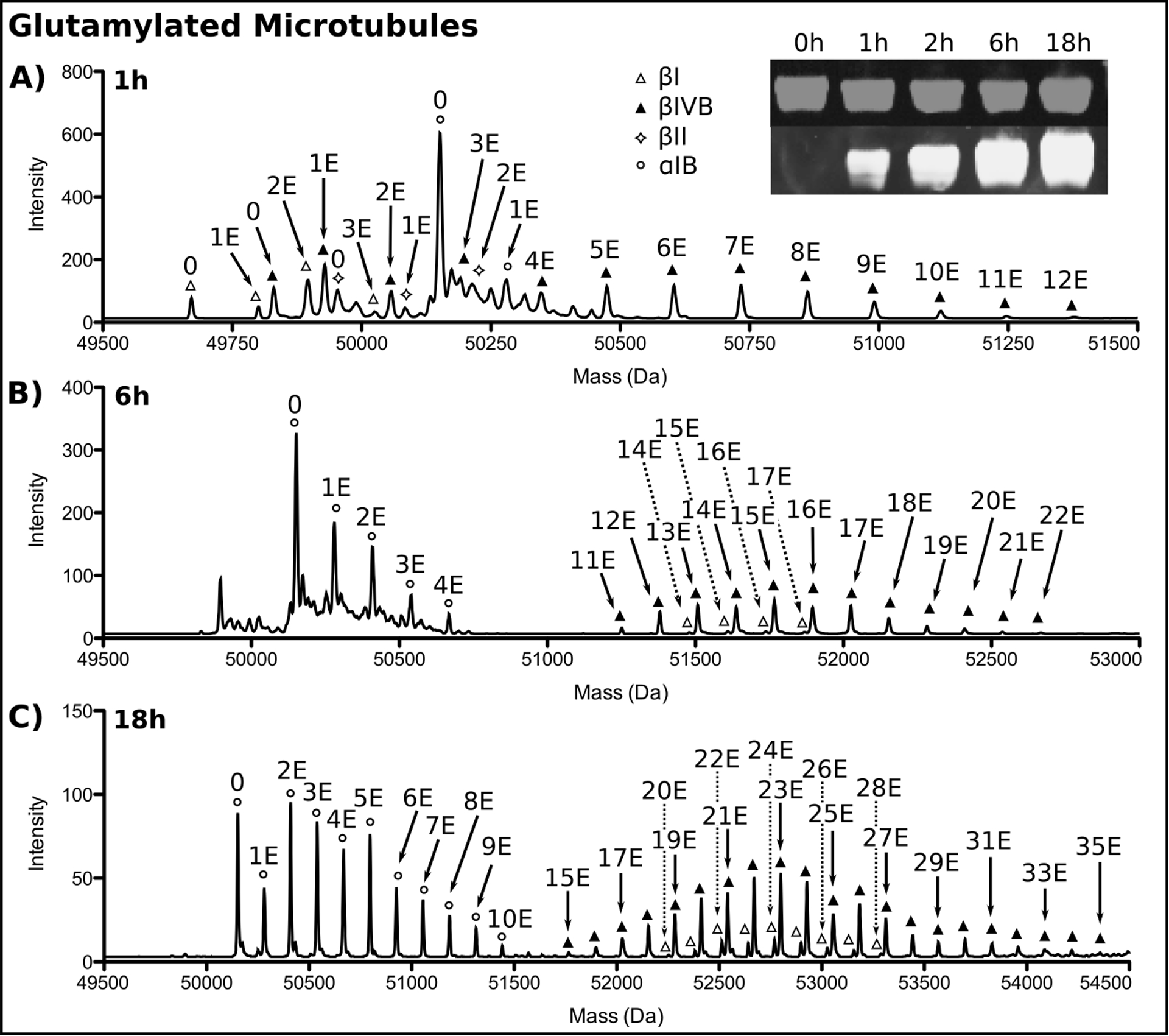
Reverse phase LC-MS and Western blot analysis of unmodified microtubules glutamylated by TTLL7. (A), 1 h, (B), 6 h, (C), 18 h incubation. Tubulin isoforms are indicated by symbols. The number of glutamates added to each isoform by TTLL7 is indicated. Only every other glutamate added to βI and βIVB at 18 h is labeled for clarity. Inset shows the progression of tubulin glutamylation monitored by Western blot at the indicated time points. α-tubulin, grey, glutamylated tubulin, white. The two channels are offset for clarity.