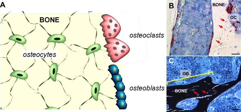
Figure 1.
A. Schematic representation of bone containing osteocytes embedded in the matrix, and osteoblasts and osteoclasts on the surface. B and C. Histologic sections of distal femur stained for B. TRAP (osteoclasts, OC, red) and counterstained with Toloduine blue and C. von Kossa (mineralized bone, black) and counterstained with McNeal. A row of osteoblasts (OB) is indicated by the yellow line. Red arrows point at osteocytes within the bone matrix. Bone sections were prepared and stained at the ICMH Histology and Histomorphometry Core, Indianapolis, IN, USA. Scale bars indicate 25μm.

