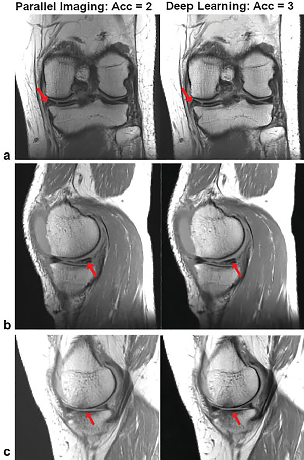
Fig. 10.
A single slice of proton-density-weighted (a) coronal and (b, c) sagittal images from 3 different patients: TR/TE, 2,800/2, turbo factor (TF): 4, matrix size: 320 × 288 (coronal) and 384 × 307 (sagittal), on a 3-T system (Skyra, Siemens) using a 15-channel knee coil. (a) A tear in the medial meniscus, (b) a complex tear of the medial meniscus and cartilage thinning with subchondral marrow changes are visible in both the parallel imaging and the variational network deep learning reconstructions.

