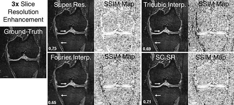
Fig. 7.
Example of knee double-echo steady-state images showing 3× resolution enhancement in the slice direction (left to right) using the deep learning super-resolution method compared with conventional techniques such as Fourier interpolation, tricubic interpolation, and a state-of the-art non-deep learning technique of sparse coding super-resolution (SC SR). The accompanying structural similarity index measure (SSIM) maps demonstrate image quality differences compared with the ground-truth image (white = high similarity; black = low similarity). The arrows indicate regions of particular image quality enhancement around the articular cartilage and the medial collateral ligament. The calculated SSIM values are displayed in the bottom left corner.

